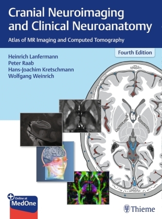
Written by experts in the field, this beautifully illustrated text/atlas provides the tools you need to directly visualize and interpret cranial CT and MR images. It reviews with exacting detail the normal anatomic brain structures identified on sagittal, coronal, and axial imaging planes. Use this book to make accurate and complete neurological assessments at the earliest possible stages - before reaching the sectioning or operating table.
\nThis revised and expanded third edition contains nearly 600 illustrations - most in color - that provide graphic representations of brain structures, arteries, arterial territories, veins, nerves and neurofunctional systems. The illustrations depict anatomic structures in shades of gray similar to the way they are seen in CT and MR images.
\nContent and illustrations expanded by more than 20%
\nHigh resolution T1 and T2 weighted MR images
\nImproved anatomic terminology for more accurate descriptions of findings
\nClinically relevant, easily readable, and clearly organized, this well-illustrated book is an essential introduction to the field for medical students and residents in neurology, neurosurgery, neuroradiology, and radiology. Practicing specialists will also benefit from this practical day-to-day tool.
ukryj opis- Wydawnictwo: Thieme, Stuttgart
- Kod:
- Rok wydania: 2019
- Język: Angielski
- Oprawa: Mieszana
- Liczba stron: 652
- Szerokość opakowania: 22 cm
- Wysokość opakowania: 31 cm
- Głębokość opakowania: 3.4 cm
- Waga: 2.3 kg


 Kontakt
 Konto
Konto


Recenzja